Historical interview: The following preserves the research experiences and personal views expressed in the original manuscript.
Research context: The device below is a historical research prototype described by the interviewee. The available material does not establish authorization for home or clinical diagnosis. Clinical care and interpretation of tests should follow the applicable clinical standards and validated testing conditions under healthcare professionals' assessment.
PhDSciNet Interview Series
A Microfluidics-Based Technology for Detecting the Coronavirus
Academic background of the science presenter:
Bachelor's degree: polymer materials science and engineering. Master's degree: biomedical engineering. Doctoral research: drug encapsulation and release. Postdoctoral research: extending applications at the interfaces of biomaterials through microfluidic technologies.
Q 1: Why did you choose this research topic?
Since its outbreak in late 2019, the coronavirus has caused immeasurable harm to society, economies, safety, and human well-being worldwide. As with other pandemics, humanity's response to the coronavirus requires accurate and effective diagnostic technologies to support efforts such as preventing outbreaks, diagnosing disease, tracing infected people's movements, interrupting transmission, and responding to viral mutations.
Q 2: What coronavirus-testing technologies are commonly used?
Coronavirus-testing technologies can be divided into several categories according to their principles:
The first category is clinical assessment, including symptoms, medical history, and, when necessary, imaging such as chest X-rays or CT, or computed tomography. These can help assess a patient's condition, but chest CT should not be used for screening or as a first-line diagnostic test for coronavirus infection.
The second category is nucleic-acid testing. The genetic material of SARS-CoV-2 is RNA; nucleic-acid amplification tests, including RT-PCR, detect specific target sequences in that viral RNA. Laboratory-based nucleic-acid tests generally have high sensitivity and are regarded as the “gold standard” in coronavirus testing, but results still need to be interpreted in light of sampling, testing conditions, and the clinical situation.
In the third category, antigen and antibody tests must be distinguished. Antigen tests look for specific viral proteins and can be used to detect current infection. Antibody tests look for the body's immune response to infection or vaccination. They do not detect the virus's “outer coat” and cannot diagnose a current coronavirus infection. The two types serve different purposes and should not be combined into a single technology with an accuracy somewhere between clinical assessment and nucleic-acid testing.
Early in the outbreak, doctors and scientists first collected clues such as clinical symptoms, considered which diseases they might resemble, and confirmed the new virus through pathogen research. After obtaining the coronavirus's RNA sequence, researchers could select suitable target sequences and design nucleic-acid tests. For other viruses, tests should likewise be designed according to their actual DNA or RNA genomes; SARS-CoV-2 itself should not be described as a DNA virus.
Once researchers have obtained a virus's genetic sequence, they can share that information to help laboratories in different places establish nucleic-acid tests. Antigen tests instead target viral proteins, while antibody tests target the body's immune response. The latter cannot replace nucleic-acid or antigen tests for detecting current infection.
Q 3: Which of these approaches is most suitable early in an outbreak?
The criteria for judging a testing technology vary between people, or rather between settings, because the best option differs across applications.
Early in an outbreak, information such as symptoms, medical history, and test records has important clinical and research value. The original interview mentioned early case studies, but this restoration does not use a particular team, journal, or citation count that has not been verified here to establish the effectiveness of a testing method.
Nucleic-acid testing plays an important role in viral diagnosis, but methods differ in operational complexity and suitability for particular settings. Patients, especially those who are severely ill, should be assessed by healthcare professionals using the clinical situation and valid diagnostic standards. This discussion should not be used to recommend an antibody test first to determine whether someone is infected.
Q 4: What are the advantages and disadvantages of these technologies?
Clinical assessment involves a doctor considering symptoms, history, and any necessary imaging. Chest X-rays and CT can show changes in the lungs, but similar changes may occur in other diseases, so those images alone cannot confirm coronavirus infection. The source's unsupported statement that diagnostic accuracy “might not even reach half” is not retained as a conclusion about accuracy.
Nucleic-acid testing often requires professional operation. RT-PCR first converts RNA into DNA, then uses amplification and signal detection to determine whether specific viral target sequences are present. Many tests need laboratory conditions, but some nucleic-acid methods can be used at the point of care; it is incorrect to say that all such tests must be performed in specialist laboratories. In settings with limited resources, equipment, electricity, staff training, and supply chains can still affect access to testing.
Antigen testing includes several specific methods. Some require instruments, while others use relatively simple formats such as lateral-flow strips. In these tests, specific antibodies are immobilized on a testing substrate to capture viral antigens in the sample, and a signal displays the result. Whether nasal swabs, saliva, or other samples can be used depends on the product's validated intended use. Antigen tests are generally more convenient than laboratory-based nucleic-acid tests, but may be less sensitive; a negative result alone cannot rule out infection. Antibody testing has different targets and purposes and should not be mixed into this process for detecting current infection.
Q 5: Could you briefly introduce the coronavirus-testing technology you developed?
Intended users: ordinary people without specialist knowledge
When we first developed the technology, our intended users were ordinary people without specialist knowledge. For example, we hoped to provide people living in less-developed parts of the world with an inexpensive coronavirus test they could operate themselves.
A few months after the outbreak, the world's major developed countries, including our own country, already had mature testing technologies based on modern molecular-biology laboratories. Although the instruments were precise and complex, those countries had substantial administrative capacity and social infrastructure, so large-scale testing was not especially difficult to implement. In many other countries, however—for example, some places in Africa—communities received insufficient effective international attention and lacked the means to develop their own technologies. Their patterns of viral transmission might not even be known. We therefore wanted to study a technology for them.
Guiding technical approach: simple operation and easily understood results
When choosing our approach, we therefore ruled out complex nucleic-acid diagnostic techniques first. We also did not want to include too many methods requiring auxiliary technologies such as fluorescence, because that would increase operational difficulty and make results harder for users to understand.
We ultimately chose the relatively direct approach of antigen testing. We wanted to develop a technology that simply detects whether a patient's body contains the coronavirus. We treat the coronavirus as the antigen, and our device contains antibodies against it. When the two bind, a readily visible signal can be presented to the user, allowing the result to be understood simply and clearly.
We designed a microfluidic chip, shown in the images, as a highly integrated, closed device. It has an inlet and an outlet. In the channel between them, we placed a small region resembling a filter mesh. Its pore size is precisely 30 micrometers: particles or viruses smaller than 30 micrometers can pass through, while particles larger than 30 micrometers cannot.
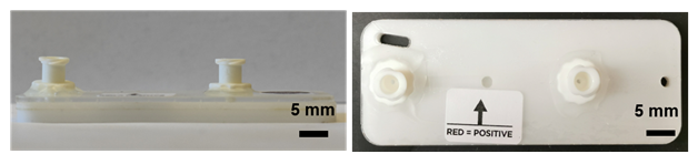
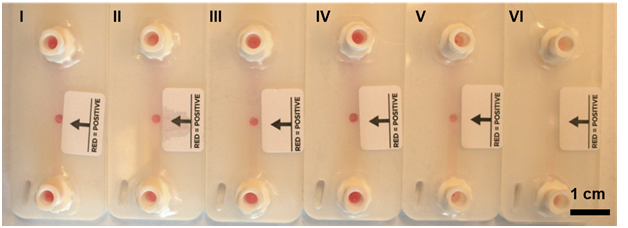
The research procedure described in the manuscript was as follows: the team prepared several milliliters of buffer for volunteers, mixed one or two drops of saliva into it, and allowed it to stand for several to several tens of minutes. If viruses in the sample reacted with particles in the buffer, the originally white microparticles could turn red. In the research process, a handheld syringe then introduced the solution into the microfluidic chip, where filtration concentrated red particles in a small area to create a visible signal. This is the interviewee's description of an experimental prototype, not instructions for readers to test themselves.
In the negative-sample experiments described by the interviewee, the particles remained white, so the designers hoped to distinguish samples through red and white signals. Actual diagnosis still requires appropriate validation: an absence of color does not directly establish that a person is uninfected.
The microparticles are made from a very simple material called polystyrene, which is also familiar as the material used for disposable white foam food containers. Their size is around 40 micrometers. We attach antibodies with high specificity for the coronavirus to their surfaces, turning them into automatic capture devices that bind the virus.
Q 6: Approximately how long does it take to obtain a result after saliva sampling?
In the laboratory, we found that the average time for color to begin developing was around five minutes. If a user was impatient, pink or pale red could appear after five minutes. But we recommended waiting thirty minutes, when the red color reached its deepest intensity.
The interviewee reported that, in their experimental observations, the red color could persist for several hours or longer, and therefore considered the display signal relatively stable. This is retained as a historical experimental observation, not a basis for clinical interpretation outside a test's valid reading window.
Q 7: How should the testing device be stored?
The interviewee explained that the prototype chip used an engineering plastic and was relatively simple to store. The buffer needed more careful storage; at the time, they recommended protecting it from light and refrigerating it without freezing. These are descriptions of research materials in the original interview. Their specific stability, shelf life, and transport conditions have not been independently verified from the available material and are not presented as current storage instructions for a testing product.
Q 8: How much does the testing device cost?
The cost depends on the engineering material you choose. Because we were making these devices in the United States, local labor and prices were relatively high. At the time, producing one chip cost us around one US dollar.
The interviewee saw a potential advantage in the prototype chip's low cost and reported trying to reuse it after flushing liquid back through the outlet, which might further reduce experimental costs. This historical device design and reuse concept do not establish validation for clinical reuse, nor do they mean that a device containing clinical samples can be rinsed with tap water and then used again for testing. It must not be treated as an operating method for a home or clinical test.
Q 9: How accurate is the testing device?
In the original interview, the presenter reported testing a batch of clinical samples in the United States “around Christmas last December.” The sample size was approximately 100, with reported accuracy of about 95% and specificity of about 100%, while emphasizing that the dataset was limited. These figures are retained as the interviewee's historical account of the research. The available material has not independently verified the statistical definitions, sample composition, or validation results, and the figures must not be treated as performance guarantees for an authorized home-testing product.
Science presenter: Eureka
Editors: Honey Peach Oolong and Calorie
Interviewer: Fantuan
Audio recording: Fantuan
Revised on 2026-10-10: Distinguished SARS-CoV-2 RNA testing, viral-antigen testing, and tests for the body's antibodies; corrected unsupported clinical statements about chest CT and test selection; limited color development, storage, and chip reuse to descriptions of a historical research prototype; and retained the interviewee's small-sample performance report with its limits and lack of independent verification here.
Supplementary references
- CDC: Overview of testing for SARS-CoV-2
- CDC: Testing for COVID-19
- FDA: COVID-19 test basics
- FDA: Uses and limitations of COVID-19 antibody tests
- American College of Radiology: Chest-imaging recommendations for suspected COVID-19
Sources and editorial history
Restored from a complete historical article exported from the PhDSciNet Official Account.
Editorial revision: Revised on 2026-10-10: Distinguished SARS-CoV-2 RNA testing, viral-antigen testing, and tests for the body's antibodies; corrected unsupported clinical statements about chest CT and test selection; limited color development, storage, and chip reuse to descriptions of a historical research prototype; and retained the interviewee's small-sample performance report with its limits and lack of independent verification here.