English translation of the original Chinese article. Publication dates and the extent of recovered text are preserved. Figures retain their original labels. Read the Chinese original.

Affiliations listed in the author's original manuscript: Shanghai Institute of Materia Medica, Chinese Academy of Sciences / East China University of Science and Technology (historical credit).
Powerful astronomical telescopes allow us to photograph the eternal beauty of the universe; cameras capture nature's strange and colorful wonders; and optical microscopes have lifted one corner of the veil over the microscopic world. However, diffraction limits make it difficult for conventional optical microscopes to resolve many nanoscale details. One nanometer is one billionth of a meter.
Our curiosity about the unknown has not faded; it has grown stronger. In 1986, G. Binnig and Ch. Gerber at IBM and Professor C. F. Quate at Stanford University developed the atomic force microscope from the scanning tunneling microscope, opening an entirely new way to observe the world. It enables nanoscale surface imaging of materials and biological samples, as well as investigation of their mechanical properties.
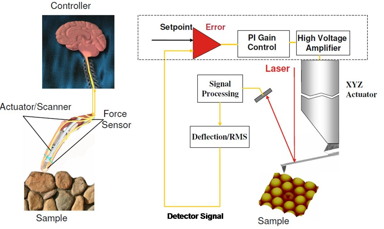
An atomic force microscope characterizes surface topography and records mechanical properties by “touching” the sample with a mechanical probe. Its operating principle (Figure 1) resembles feeling an object's surface with a finger. As the probe approaches the surface, an interaction force arises between them, deflecting the cantilever. A laser diode produces a beam that a lens focuses on the back of the cantilever, from which it is reflected onto a photodiode. During scanning, a feedback-control system regulates the microcantilever as it bends and moves over the surface topography. The reflected beam shifts accordingly, allowing the detector to record topographic and mechanical information.
Under suitable conditions, AFM can resolve molecular-scale structures. A 2013 study observed intermolecular contrast in an 8-hydroxyquinoline system at positions consistent with possible hydrogen bonds (Figure 2). Hydrogen bonding is a type of attractive interaction involving hydrogen atoms, often between hydrogen bonded to atoms such as oxygen or nitrogen and neighboring atoms.
However, line-like features in an image do not amount to directly “seeing hydrogen bonds.” Follow-up experiments in 2014 showed similar contrast where no intermolecular bonds should exist. Flexibility at the probe tip is important in explaining this phenomenon; interpretation requires structural models and other evidence.[2]
![Figure 2: AFM images and structural models of 8-hydroxyquinoline assemblies (original manuscript illustration; see reference [1]).](/assets/local/afm-local-figure-2-86ac437e69.webp)
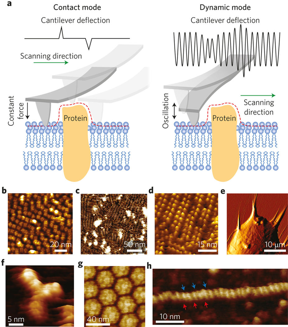
Biologists also use AFM to investigate membrane-protein arrangements, interactions between cells and fibers, antibody and virus shapes, and DNA structures (Figure 3). Different imaging modes suit different samples and have distinct limits in resolution and interpretation.[3]
As AFM scanning becomes more precise and is combined with other spectroscopic characterization instruments, scientists will reveal still more wonders of the nanoworld.
References for Restoration and Correction
- Real-Space Identification of Intermolecular Bonding with Atomic Force Microscopy. Science (2013).
- Intermolecular contrast in atomic force microscopy images without intermolecular bonds. Physical Review Letters 113, 186102 (2014).
- Dufrêne et al. Imaging modes of atomic force microscopy for application in molecular and cell biology. Nature Nanotechnology 12, 295–307 (2017).
The text was recovered from a local Word manuscript of the same work. The final text published on the original site has not yet been obtained for comparison, so this version may differ from the one published at the time. The historical date and original title are retained from the original site's index.
Editorial note: On October 10, 2026, corrected the statement directly equating line-like intermolecular AFM contrast with hydrogen bonds, and added explanations of nanoscale dimensions, figure captions and references. The author credit was restored from the manuscript of the same work.