
English translation of the original Chinese article. Publication dates and the extent of recovered text are preserved. Figures retain their original labels. Read the Chinese original.

Inside our eyeball is a transparent, jelly-like substance called the “vitreous” [1]. Behind the vitreous is a structure called the “retina.” It detects light entering the eye and sends the information to the brain, enabling us to see the outside world [1].
As we age, collagen fibers in the vitreous can clump together and cast shadows on the retina [3]. When light enters the eye and is blocked by these small clumps, we see gray or black objects drifting across our field of vision [3]. These are called “floaters,” because they look a little like small insects flying around. Floaters can occur at different ages, but a sudden appearance of many new floaters, flashes of light or an obstruction in the visual field warrants prompt assessment by an eye specialist. In older eyes, the vitreous starts to become more liquid and shrink, eventually separating from the retina [1, 3]. This is called “posterior vitreous detachment.” In most people, the separation causes no problems. In a minority, however, the vitreous pulls on the retina, tearing it and creating a small hole [1, 3]. Fluid entering through the hole may cause the entire retina to detach. Retinal detachment can occur at any age but most commonly develops after the age of forty [1, 2].
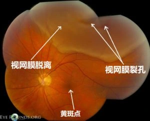
A photograph of an affected retina
Retinal detachment is a serious condition requiring eye assessment and treatment as soon as possible. The timing of treatment depends on factors such as the extent and location of the detachment; delay may cause permanent vision damage. There are four symptoms to watch for: (1) severe floaters, as though many small black insects were falling together; (2) flashes of light in the visual field; (3) a dark curtain appearing to fall across the visual field, possibly from any direction—above, below, left or right; and (4) deteriorating vision [3, 4, 8]. If you have any of these symptoms, you must seek an eye examination immediately, because this is an emergency. The doctor will dilate your pupils and examine the back of the eye. Depending on the particular tear or detachment, the doctor may arrange laser treatment, cryotherapy or surgery [4]. Prompt treatment helps protect vision, but the degree of recovery varies between individuals. Delay may increase the risk of permanent vision damage.
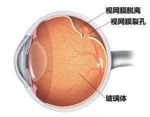
Schematic illustration of retinal disease
People with high myopia (-6D or more myopic, equivalent to over 600 on the Chinese prescription “degree” scale) must pay particular attention to the symptoms of retinal detachment [1, 4, 6]. As myopia becomes more severe, the eyeball elongates further, stretching the retina [4, 5]. The retina becomes progressively thinner, potentially developing a tear and detachment [4, 5]. Eye inflammation, eye surgery, proliferative diabetic retinopathy—an eye disease associated with diabetes—family history, lattice degeneration—thinning at the retinal periphery—and eye trauma may also lead to retinal detachment, so its symptoms require special attention [1]. The risk of retinal detachment after previous cataract surgery requires long-term attention [7].
References
[1] Dahl, A. A. & Stöppler, M.C. (2017, October 13). Retinal Detachment. Retrieved from https://www.medicinenet.com/retinal_detachment/article.htm
[2] National Eye Institute. (2009, October). Facts About Retinal Detachment. Retrieved from https://nei.nih.gov/health/retinaldetach/retinaldetach
[3] WebMD. (2017). Eye Floaters: Causes, Symptoms, and Treatment. Retrieved from https://www.webmd.com/eye-health/benign-eye-floaters#1
[4] Lai, T.Y.Y. (2007, September). Retinal Complications of High Myopia. The Hong Kong Medical Diary. 12(9), 18-20. Retrieved from http://www.fmshk.org/database/articles/07mb05.pdf
[5] Curtin, B. J., & Karlin, D. B. (1970). Axial length measurements and fundus changes of the myopic eye. I. The posterior fundus. Transactions of the American Ophthalmological Society, 68, 312–334. Retrieved from https://www.ncbi.nlm.nih.gov/pmc/articles/PMC1310382/
[6] The Eye Disease Case-Control Study Group. (1993, April 1). Risk Factors for Idiopathic Rhegmatogenous Retinal Detachment, American Journal of Epidemiology,137(7), 749–757, https://doi.org/10.1093/oxfordjournals.aje.a116735
[7] Erie, J. C., Raecker, M. E., Baratz, K. H., Schleck, C. D., & Robertson, D. M. (2006, December). Risk of retinal detachment after cataract extraction, 1980-2004: a population-based study. Transactions of the American Ophthalmological Society, 104, 167–175. Retrieved from https://www.ncbi.nlm.nih.gov/pmc/articles/PMC1809901/
[8] Montague, C & Vislisel, J. (2016). This photograph shows a superior rhegmatogenous retinal detachment that is not yet involving the macula [Fundus Photograph, Labels Added]. Retrieved from https://webeye.ophth.uiowa.edu/eyeforum/atlas/pages/Rhegmatogenous-ret-detach.htm
[9] Mayo Foundation for Medical Education and Research. (2017). Retinal Detachment [Multimedia image, edited]. Retrieved from https://www.mayoclinic.org/diseases-conditions/retinal-detachment/multimedia/retinal-detachment/img-20006758.
Additional References
- NIH National Eye Institute: surgery for retinal detachment and the timing of treatment
- NIH National Eye Institute: laser treatment and cryotherapy for retinal tears
The text was recovered from the matching article retained by the WeChat account, with the original site's publication record preserved. The old WeChat promotional layout has been removed, and available original illustrations have been restored.
Editorial note: Restoration and correction on 2026-10-10: corrected the mechanism of floaters and removed absolute statements about a fixed surgical deadline and recovery of vision. The article continues to make clear that retinal detachment is an emergency requiring prompt care.