
Historical interview: the following preserves the research experiences and personal views expressed in the original manuscript.
3D bioprinting is an emerging technology that uses a computer-generated three-dimensional model as a blueprint and specially prepared “bioink” to manufacture artificial organs and biomedical products. As an interdisciplinary frontier technology, it has given further momentum to the rapid development of regenerative medicine.
3D bioprinting
What specific applications does 3D bioprinting have?
Orange Soda: in academia, people usually apply 3D bioprinting to tissue engineering, such as printing organs or tissues. One main research issue is finding suitable printing materials. Developing and applying bioinks is actually central to bioprinting. Our aim is to develop a stable bioink that balances printability, biocompatibility, mechanical properties, and other requirements.
If I want to print the structure of a heart, liver, or blood vessel, for example, I design the ink according to its properties, such as mechanical strength and electrical conductivity, then print the desired structure. I can also mix cells into the bioink and examine how they grow in the printed structure and whether they function as intended. If their function is poor, the bioink is not particularly good and needs further improvement.

What businesses do commercial bioprinting companies mainly pursue?
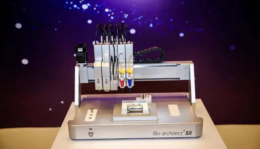
Orange Soda: they mainly follow two directions. One is making 3D printers. Existing plastic printers can be adapted for biological use: where early machines had one nozzle, four- or five-nozzle printers are now common, and more elaborate versions resemble robotic arms. If a structure needs more materials, different bioinks can be stored in something like a bank. During printing, the arm collects the appropriate cartridge, prints with it, then collects another material. This allows as many printing materials as needed.
The other direction is developing bioinks. Common materials include gelatin and hyaluronic acid, which are very ordinary and familiar. Companies hold relevant patents, use different material ratios, or add other components.
Can 3D bioprinting produce human skin?
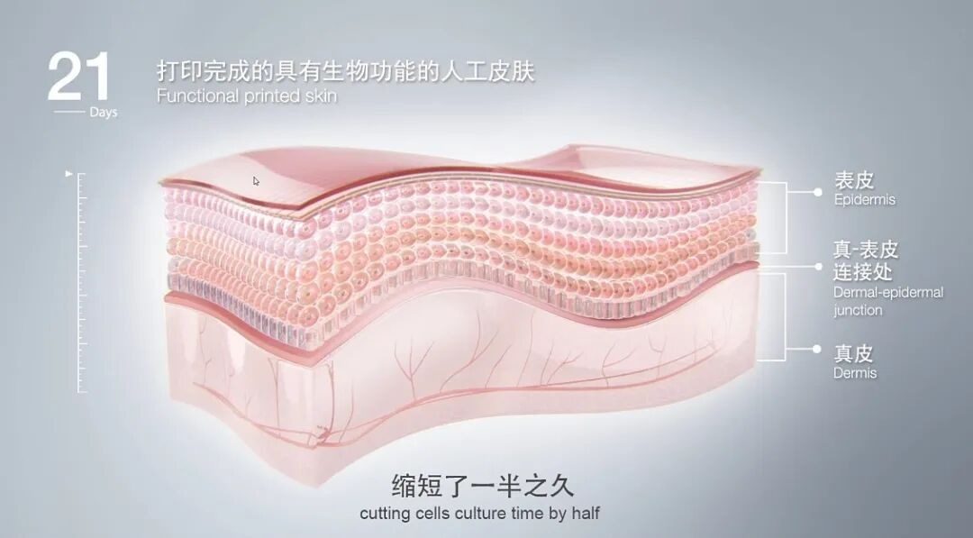
Orange Soda: skin is actually the easiest to print because its structure is not particularly complex, and people began researching it several years ago. Appropriate bioinks are prepared for the different layers of skin and printed one layer at a time.
For example, if someone has a burn, you can print skin matching its shape. Scanning the wound first produces an image, which software converts into a three-dimensional design. Feeding that into the printing software lets the machine print a layer representing the skin structure.
Can 3D bioprinting produce bone?
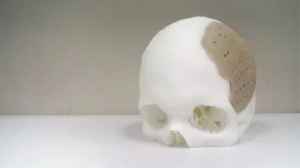
Orange Soda: yes, work is being done on bone, and bone-related products have already reached the market. Repairing damaged bone commonly uses artificial bone, such as titanium alloys. There is also 3D printing of inorganic materials to make three-dimensional bone structures.
A Chinese company, Guangzhou Medprin Regenerative Medical Technologies Co., Ltd., has produced bioprinted dura mater patches that have been implanted in the brains of more than ten thousand patients.
Technical content check: nonliving 3D-printed orthopedic implants and dural patches differ from bioprinting organs containing living cells. Printing an organ shape does not establish a fully functional organ suitable for transplantation; the FDA describes printing living hearts, livers, and similar organs as research-stage work. The patient count is a historical interview statement and has not been verified as current data.
3D bioprinting and organ transplantation
Why could 3D bioprinting be used for organ transplantation?
Orange Soda: organ transplantation may be what people first think of when discussing bioprinting. It seems promising because printing materials can come directly from a patient: cells are cultured and mixed with biomaterials to produce the bioink needed for organ printing.
If an organ has problems and needs replacement, we can print its required shape and culture it outside the body. If the biomaterials are all optimal, the patient’s cells placed within them can grow in the printed structure and generate their own tissue. The external biomaterials are not part of the human body but are designed to degrade. If the rate at which cells produce extracellular matrix matches the degradation rate of the original printing material, an organ closely matched to that patient may ultimately grow.
Why not simply let cells grow on their own? Mixing them with a biomaterial and printing the structure guides them to grow into something suited to the patient’s body. That is the ideal situation.
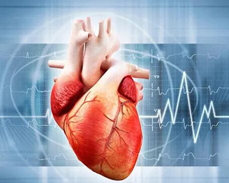
Applications of bioprinting in drug research
Orange Soda: pharmaceutical development requires two-dimensional experiments, animal experiments, and clinical trials, progressing step by step before a product can be marketed for patients.
That normally takes a very long time. Bioprinting provides a three-dimensional environment that simulates the human body and can simplify the process. Suppose a drug treats a lung disease. If we print tissues representing a lung, heart, and liver and put all three on a chip, administering the drug lets us observe effects on the heart and liver at the same time. We can study its metabolism in liver tissue and infer possible effects on human health while also examining whether it acts on the lung. This can be more humane than traditional drug experiments.
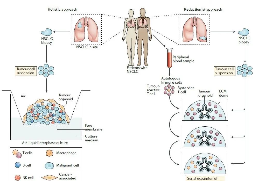
How long does it take to bioprint a human organ?
Orange Soda: printing time depends on the organ’s volume. The main time-consuming part is subsequent cell growth and culture.
Current research mainly concerns drug screening, where requirements are less strict. If I only need to see whether a drug affects liver cells, for example, results may be obtained within three or four weeks without waiting for an actual liver to develop.
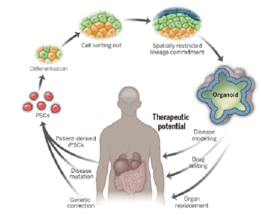
Do different organs require different combinations of bioinks?
Orange Soda: yes. Different organs and tissues have different mechanical strengths and functions, so ideally we need different materials for different parts to simulate a human organ properly.
Could bioinks cause rejection if printed tissues or organs are transplanted into a person?
Orange Soda: a printed organ must first be cultured outside the body before transplantation. During culture, cells develop tissue while the original bioink degrades. Rejection mainly results from differences in human leukocyte antigens between recipient and graft. I therefore do not think the problem is rejection of the material: if it does not cause inflammation, it should not cause rejection when used in the patient.
The organ is printed using the patient’s own cells, so transplanting their own material back into them should not cause problems. If other inflammation occurs, that indicates the bioink is not ideal and needs further research.
Medical content check: using a patient’s own cells does not guarantee that an implant has no immune response or other risks. Biomaterials and scaffolds can still cause inflammation, fibrosis, and related responses; safety and function require specific research and clinical evaluation.
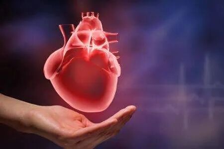
A bioprinting challenge: printing blood vessels
What is the greatest challenge when bioprinting human organs?
Orange Soda: organ structures can currently be printed, but their function is incomplete. A heart, for example, contains many blood vessels, and printing those vessels is a current challenge.
The body requires many blood vessels. Without them, as a printed structure becomes more complex, nutrients cannot reach its center adequately and cells have difficulty surviving there. Printing many vascular structures within the tissue is therefore a major challenge under active investigation.

So the key issue is how to print blood vessels?
Orange Soda: yes. Once a structure is more than one hundred to two hundred micrometers thick, the cells’ nutrient supply is already quite limited. Blood vessels supply nutrients to cells within that surrounding distance, helping them survive and function. Without vessels, cells in thicker central areas receive insufficient nutrients and may die or function poorly.
Does printing a vessel involve putting vessel-wall cells in a mold and allowing them to grow?
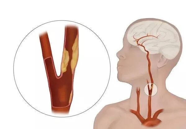
Orange Soda: some can be printed directly. The carotid artery, for example, is relatively large, with an internal diameter reaching six millimeters, much larger than capillaries or some other vessels, so it can be printed conventionally. A supporting bath can hold the structure during printing, allowing tall, large forms without collapse.
That applies to larger structures. For smaller ones, one approach is coaxial, two-component printing: a small nozzle sits inside a slightly wider one. One solution runs through the inner nozzle and another through the outer one. The inner solution cross-links the outer material, directly producing a hollow tube resembling a vessel. A simple example uses alginate solution outside and calcium chloride inside: contact cross-links the alginate into a hydrogel. Controlling the relative extrusion speeds produces the vessel structure.
At the current pace, how long might it take to solve this challenge?
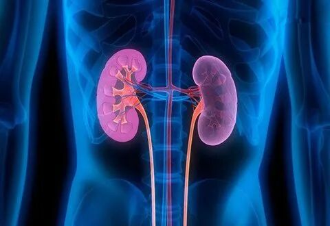
Orange Soda: I have seen Professor Anthony Atala’s group, which also studies bioprinting, print a kidney, perhaps around 2010 or 2011. But there is still a long way to go before actual clinical use. Without solving the vascular problem, printed organs cannot be used for human transplantation.
Some questions about 3D bioprinting
What parameters matter when choosing printing materials?
Orange Soda: one is mechanical strength. For a liver, the biomaterial’s strength should match that of a human liver, providing a relatively friendly environment for liver cells. That comes first.
Second, as a biomaterial, it must be nontoxic and biocompatible, and we must consider its degradation rate.
Third, for extrusion-based printing, we must consider shear-thinning behavior: viscosity is low at a high shear rate and high at a low shear rate. Cells experience squeezing during printing. If shear stress—shear rate multiplied by viscosity—is too high, cells die because they are relatively fragile. High viscosity at a low shear rate means that after extrusion the ink does not flow easily and holds its shape, improving structural stability.
How accurate is the information from bioprinting-based drug screening?
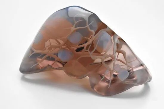
Orange Soda: if I remember correctly, older methods used two-dimensional tests before animal or clinical trials. Cells were tested in a flat environment, but their behavior there differs from behavior in the three-dimensional human body.
Traditional methods may therefore already be inaccurate at that stage. Better simulation of the human environment should improve accuracy and cellular function, although I think clinical trials will certainly still be required before marketing. The earlier stage becomes more accurate: compared with conventional two-dimensional screening, cells in a three-dimensional environment should provide more accurate results.
Are there ethical issues in applications of 3D bioprinting?
Orange Soda: bioprinting creates an object without changing human gene sequences or violating the principles of life. If developed well, it could meet society’s need for organs, so ethical issues are not discussed very much. Currently it mainly serves drug screening, and I think good work there would be very beneficial.
Ethics content check: leaving gene sequences unchanged does not remove ethical questions. Research involving human cells, tissues, or participants must still consider informed consent, participant protection, and safety; these principles also apply to organ-printing research.
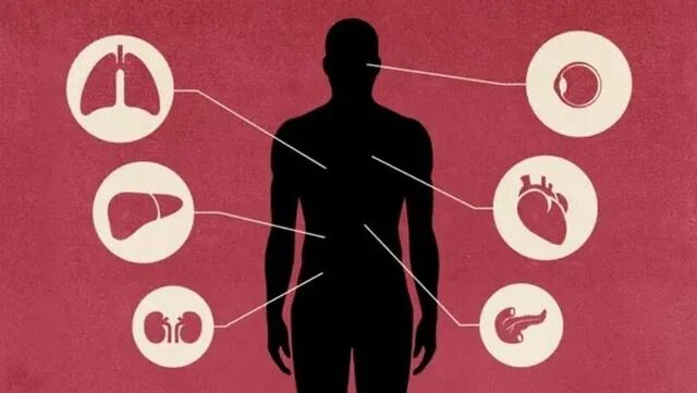
In theory, bioprinting can construct skin, bone, nerves, organs, and other structures outside the body. The complexity of human organ structure, shape, and function still poses many challenges, but greater understanding of multicellular, multimaterial, highly complex systems should accelerate regenerative medicine as bioprinting improves, bringing revolutionary advances to modern medicine.
Science contributor: Orange Soda
Text editors: Fantuan, Calorie
Audio editor: Honey Peach Oolong
Interview: Fantuan, Calorie
Audio recording: Honey Peach Oolong
This article expresses the author’s personal views and does not represent those of this website. Images were sourced online; please contact us regarding any infringement.
Additional sources checked
US Food and Drug Administration: Medical applications of 3D printing
US National Institutes of Health: Tissue bioprinting and drug research
US National Institute of Biomedical Imaging and Bioengineering: Immunoengineering
Wake Forest University: Anthony Atala profile
US Office for Human Research Protections: Research guidance
Sources and editorial history
Restored from a complete historical article exported from the PhDSciNet Official Account.
Editorial revision: The historical interview and research outlook are retained. Anthony Atala’s name was corrected, with checks distinguishing medical implants from living-organ printing, risks associated with autologous cells, and human-research ethics.