English translation of the original Chinese article. Publication dates and the extent of recovered text are preserved. Figures retain their original labels. Read the Chinese original.
Li Ka Shing Knowledge Institute, St. Michael’s Hospital, Toronto, Canada
Keywords: microfluidics, cancer detection, cell sieve
Introduction
Cancer inspires fear because of three characteristics: uncontrolled proliferation, a tendency to mutate readily, and the ability to metastasize. Advanced cancer often comes with serious complications throughout the body, causing patients great suffering and frequently leaving modern medicine with few effective options. Treating and controlling early-stage cancer, however, is relatively much easier. Early detection and timely treatment can greatly improve patients’ five- to ten-year survival rates and may even achieve a complete cure.
Detecting cancer early, however, is a difficult problem. In films and television programs, we often see doctors examining a patient’s tissue sections under a microscope; such scenes are not much of an exaggeration. Cancer diagnosis usually combines medical history, examination, imaging, laboratory tests and, when needed, a tissue biopsy. Biopsy is an important basis for diagnosis in many situations, but this does not mean that any single test can be described as diagnosing cancer with “100% certainty.” Sampling also has limitations, including its invasive nature. Is there a way to investigate early cancer quickly, with minimal invasion and accurately?
There are approaches worth investigating. As noted above, one troublesome feature of cancer cells is their tendency to spread. Some particular surface proteins can make them more mobile than normal cells [1], allowing migration through lymph, tissue fluid and blood. Cells released into the bloodstream are known as circulating tumor cells, or CTCs. CTCs are relevant to research on tumor dissemination and prognosis, but a result must be interpreted alongside other clinical information. A single CTC test cannot simply be equated with a cancer diagnosis.
A further question arises: are CTCs easy to detect? This is difficult for conventional methods. Although typical cancer-cell sizes described here (14–26 micrometers) exceed those of normal red blood cells (6–9 micrometers) and white blood cells (8–20 micrometers), and they have distinctive morphological features [2], CTCs are very rare in human blood. Separating, identifying and counting them among large numbers of normal blood cells is challenging, so researchers need specialized enrichment and detection methods. Are other highly sensitive approaches available? Yes: the microfluidic technology investigated for early cancer detection that this article introduces.
An Introduction to Microfluidics
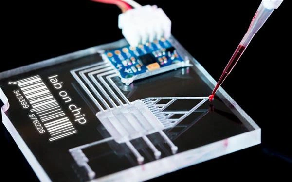
Microfluidics is a technology for controlling, manipulating and analysing complex fluids at microscopic scales. It emerged in the early 1990s as an interdisciplinary field combining physics, chemistry, biology, medicine and manufacturing engineering. As the name suggests, “micro” refers to small dimensions and high precision: fluids can be observed and manipulated over scales from a few micrometres to several hundred micrometres. The “fluid” being studied may be a solution, blood, a gas or even a supercritical fluid. Modern microfluidic technology can integrate hundreds or even tens of thousands of functional units on a glass or plastic chip substrate covering only a few square centimetres. With a simple experimental design, researchers can manipulate anywhere from a few to hundreds of millions of basic microscopic units, such as tiny droplets, bubbles and biological cells, as shown in Figure 1.
Figure 1: A polymer-based microfluidic chip for blood analysis and testing (source: Brigham Young University, United States).
A Microfluidic “Cell Sieve”
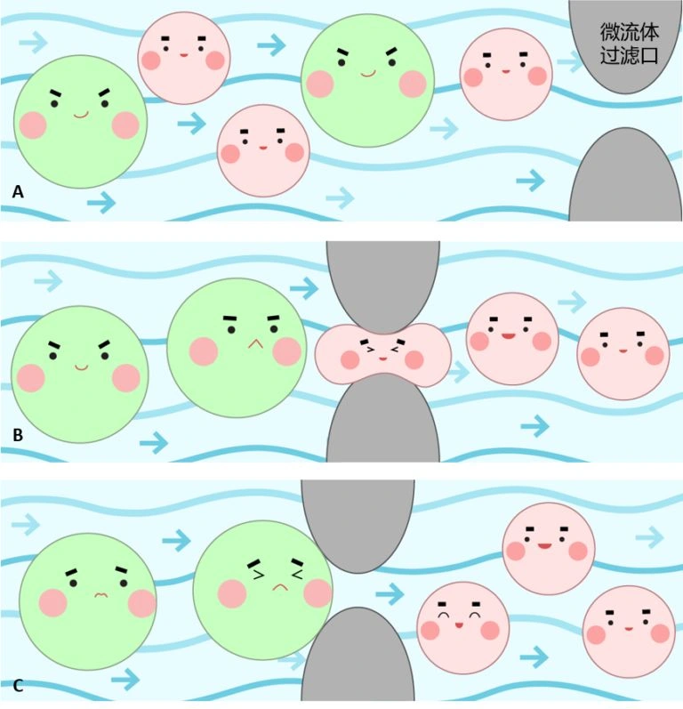
First, a small microfluidic chip is prepared from the polymer polydimethylsiloxane (PDMS; see Figure 1). Biocompatibility, cleaning and conditions for reuse must be validated for the particular device. Cancer cells and normal cells enter the microfluidic channels together. The far end resembles a sieve, with openings somewhat larger than normal blood cells but smaller than the cancer cells, acting as a filter. As the mixture passes through, cancer cells are retained while red blood cells and most white blood cells pass freely. A minority of larger white blood cells can also pass under appropriate pressure because they are soft and deformable. This enriches larger cells from the blood. Immunolabeling, cell morphology and other analyses are then needed to identify CTCs; filtering by size alone does not diagnose cancer (see Figure 2).
Figure 2: (A) Cancer cells (green) and blood cells (red) are filtered together in a microfluidic channel; (B) blood cells pass normally through the filtering channel; (C) cancer cells cannot pass through it. Illustration by 罗楠, UX designer, Finastra, Toronto.
Sample volumes and performance vary with the experimental design of each microfluidic platform. The study cited in the original article [3] reported recovery rates of 68%–100% for different cultured lung-cancer cell lines spiked into blood. Its clinical investigation involved patients already diagnosed with advanced lung cancer. These data cannot be treated as the accuracy of early cancer screening in the general population, nor can a cell-capture rate be directly equated with the accuracy of cancer diagnosis.
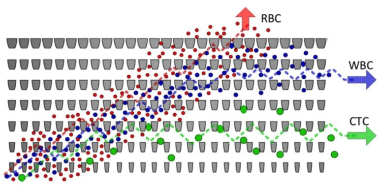
Figure 3: A schematic of an array of microfluidic filtering channels. RBCs (red blood cells), WBCs (white blood cells) and CTCs (circulating tumour cells) are separated into different channel outlets for collection and analysis [4].
Conclusion
The method is simple, fast, efficient and convenient. Even the tiny numbers of circulating tumour cells in blood have nowhere to hide in the microfluidic “net”: we can quickly capture and analyse them one by one. This is precisely the advantage of microfluidics over conventional detection techniques. Many pharmaceutical companies and research institutions are vigorously advancing large-scale commercial applications of this technology. Microfluidic detection is rapidly finding its way into, and assisting, many aspects of cancer research and treatment, and we can expect to benefit more from it in the near future. Of course, we hope everyone eats healthily, stays physically active and avoids cancer altogether!
*This article expresses the author’s personal views, not those of this website. Other media outlets, websites or individuals who reproduce it must credit the source and bear their own responsibility for copyright and other legal matters. Authors who do not wish their work to be reproduced, or who wish to discuss reproduction fees, should contact us.
References:[1] Balic M, Williams A, Lin H, et al. Circulating tumor cells: from bench to bedside. Annu Rev Med, 2013, 64: 31-44
[2] Gorges T M, Tinhofer I, Drosch M, et al. Circulating tumour cells escape from EpCAM-based detection due to epithelial-tomesenchymal transition. BMC Cancer, 2012, 12(1): 178
[3] Hosokawa M, Kenmotsu H, Koh Y, et al. Size-based isolation of circulating tumor cells in lung cancer patients using a microcavity array system. PloS One, 2013, 8(6): e67466
[4] Tilman Todenhöfer et al.,Urologic Oncology,Volume 34, Issue 11, Pages 483.e9–483.e16
Images from the same-title WeChat manuscript
The following images come from the preserved WeChat manuscript with the same title and have been restored in their original order.
Sources for this correction: National Cancer Institute · How Cancer Is Diagnosed; NCI · Circulating tumor cell; Hosokawa et al., PLOS ONE (2013).
Additional illustrations have been recovered from the preserved WeChat manuscript with the same title and are grouped after the article. The archived text and existing editorial corrections have been retained.
Editorial note: On October 10, 2026, the relationship between CTC detection and cancer diagnosis was corrected using NCI guidance and the original paper. Experimental cell recovery was distinguished from clinical screening accuracy, and unverified absolute safety and quantity claims were removed. The original author, date and figures are retained.