
English translation of the original Chinese article. Publication dates and the extent of recovered text are preserved. Figures retain their original labels. Read the Chinese original.
Shenzhen Institutes of Advanced Technology, Chinese Academy of Sciences / MIT McGovern Joint Institute for Brain Cognition and Brain Disorders
Keywords: emotions, learning ability, optogenetics
Many readers have probably watched Discovery Channel’s reality television program Man vs. Wild. In each episode, British adventurer Bear Grylls enters environments unsuitable for human survival, such as deserts, swamps, forests, and canyons, to simulate how to escape extremely harsh conditions. In the program, Bear catches larvae, scorpions, and other creatures in the wild and eats them raw, while also demonstrating ways to survive in the desert.
But do difficult circumstances in everyday life really possess such a power, enabling those who encounter them to move forward through adversity?
Figure 1: A poster for the reality television program Man vs. Wild.
Pressures and stressors from various sources are things that species must face during evolution, and that we humans cannot avoid in our everyday work and lives.
Everyone hopes to stay away from pressure and from realities they would rather not accept. But to survive or make progress, we must bravely face some forms of “adversity.”
“Blooming alone against the cold” describes the resilience of plum blossoms. The quality used to measure someone’s ability to handle problems in adversity is called the Adversity Quotient, and is also described as an inner foundation for success.
While facing “adversity,” a positive “mindset” in the brain can not only help people overcome it successfully but also enhance the brain’s learning ability to a considerable degree.
So, what secrets lie within the complex neural networks of our brains that allow us to face “adversity” calmly and strengthen our ability to adapt to the environment, learn, and remember?
Are Positive or Negative Emotions the Key to Survival?In 1972, the American psychologist Paul Ekman proposed the hypothesis of six basic human emotions: fear, joy, sadness, disgust, anger, and surprise. These can also be classified by their nature as positive or negative emotions. How do changes in our emotions affect cognitive behavior?
Figure 2: Different emotions.
In an experiment, Australian researchers induced positive or negative emotions by asking participants to watch films and recall happy or sad events. They then asked the participants to judge whether rumors were true.
The results showed that, compared with people in a happy mood, those in a low mood were less impulsive and less likely to believe rumors readily.
We already know that, under normal circumstances, the brain’s reward centers respond to natural rewarding stimuli and positive expectations.
This suggests that positive emotions and expectations activate reward circuits in the brain, encouraging people to make positive predictions about future goals and thereby increasing their motivation to act.
Positive emotions can also stimulate creativity, adaptability, and self-confidence, helping individuals maintain the motivation to move forward even in adversity. Pessimistic emotions, by contrast, lead us to make negative predictions about future goals, reduce our motivation to act, and make us more likely to become discouraged or even give up.
The relevant neural circuits and mechanisms, however, remain unclear. Because of the particular nature of human research participants, the two studies described above could not examine these effects in detail at the circuit or cellular level.
The teams of 王建枝 at Tongji Medical College, Huazhong University of Science and Technology, 王立平 at the Shenzhen Institutes of Advanced Technology, Chinese Academy of Sciences, and 徐富强 at the Chinese Academy of Sciences’ Institute of Physics and Mathematics collaborated, pooling resources and their respective technical strengths. They discovered a previously unknown neural pathway through which emotions affect spatial learning and memory, and found that positive and negative emotions could respectively regulate animals’ spatial learning ability in opposite directions through this pathway.
What happens when this pathway is activated or inhibited? What are the specific cellular and molecular mechanisms involved? Could these findings help us develop a new generation of diagnostic techniques and treatments for disorders involving declining working memory, such as Alzheimer’s disease? All these questions require a deeper understanding.
The Hippocampus and Memory vs. the Amygdala and EmotionBefore exploring these questions, we need to understand the relationship between the hippocampus and memory, and between the amygdala and emotion.
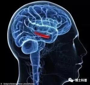
The Hippocampus and MemoryThe hippocampus is an important component of the brain’s limbic system. Named for its resemblance to a seahorse, it is primarily responsible for learning and memory.
Figure 3: The hippocampus is shown in red.
The hippocampus is responsible for the working memory an individual is currently using, such as learning new words. It is somewhat like a computer’s working memory, temporarily retaining short-term memories for hours or days so they can be accessed quickly. Short-term memories from everyday life are stored in the hippocampus. If a memory fragment, such as a word or an event, is repeated over a short period, the hippocampus transfers it to the cerebral cortex, where it becomes a long-term memory.
Damage to the hippocampus causes memory impairment, as in Alzheimer’s disease, commonly referred to in Chinese as senile dementia.
Over the past several decades, the hippocampus has been widely studied as a major brain region involved in spatial memory and navigation, using rodents as research subjects.
Researchers have found that certain hippocampal neurons become active, firing at high frequencies, only when an animal reaches a particular location. These neurons are called place cells. Animals obtain information about external features through their various senses. Place cells work with other cells in the hippocampus to compare these incoming features with previously recorded features of different locations. When the information matches, the specific place cells corresponding to that location become active.
In this way, our brains link particular features to particular spatial locations, forming memories of spatial positions.
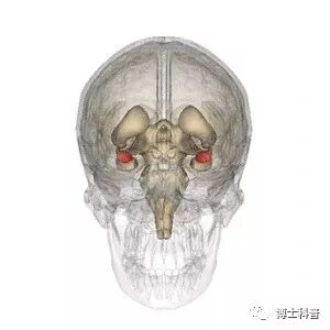
The Amygdala and EmotionThe amygdala is an almond-shaped structure attached to the end of the hippocampus, and is another important region of the limbic system.
Figure 4: The amygdala is shown in red (image source: Tavistock Institude China).
The amygdala is a brain center for generating, recognizing, and regulating emotions, and a major region controlling emotional learning and memory. Stimuli with emotional significance evoke strong electrical responses in the amygdala and form lasting traces stored in the brain.
Events that provoke strong emotional responses therefore leave long-lasting, sometimes lifelong, memories.
When amygdala function is normal, mice are naturally afraid of cats. Similarly, when someone shouts that a wolf is coming, others run away or pick up weapons to fight. If the amygdala is damaged, however, individuals may lose their fear response or even become emotionally indifferent.
In recent years, substantial research has examined how amygdala subregions and their neuronal subtypes modulate negative emotions, including anxiety, depression, and fear.
Mice “Surviving Adversity”How emotional states in humans and animals affect cognitive behavior is a widely discussed public topic.
At present, most laboratories use animal models of depression, anxiety, and similar conditions to study how negative emotions affect cognitive behavior. For example, experimental manipulations induce depressive symptoms in animals, after which researchers detect impaired memory.
However, an animal model that remains optimistic under stress is still lacking for studying how positive emotions change cognitive behavior.
In this study, 王建枝’s team established, for the first time, an animal model that retained a sense of hope under stress (learned hopefulness, LHF).
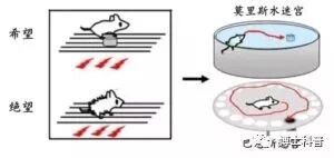
The researchers delivered an aversive stimulus to mice through foot shocks. An insulated platform was placed within the shock setting. Through repeated guidance and training, the mice learned to actively seek out and climb onto the platform, successfully ending or avoiding the foot shocks.
Conversely, when the platform was removed, repeated inescapable foot shocks significantly induced emotional responses of despair in the mice, thereby establishing an animal model of despair (learned helplessness, LHL).
Figure 5: The “hope” group had an insulated escape platform in the foot-shock setting, while the “despair” group was trapped with nowhere to escape. After several days of training, two classic mazes were used to test spatial learning ability. (Image modified; source: Nature.)
Control animals were placed in the same setting but received no electrical stimulation.
The experimenters used two behavioral paradigms—the Morris water maze and the Barnes maze—to test differences in spatial learning ability among these three groups.
The team unexpectedly found that, compared with the control group, the “hopeful” mice found an escape location more quickly, reducing the time required by about two-thirds, while the “despairing” mice showed the opposite pattern: the delay before successfully finding shelter almost doubled.
These data showed that “hope” effectively promoted spatial learning and memory in mice, whereas “despair” markedly impaired these abilities.
Discovering and Regulating the “Pathway”The teams of 王立平 at the Shenzhen Institutes of Advanced Technology, part of the Chinese Academy of Sciences’ Center for Excellence in Brain Science, and 徐富强 at the Wuhan Institute of Physics and Mathematics investigated the workings of neural circuits in the animals’ brains. Combining their technical strengths, they resolved the structure and function of this circuit. First, they used a distinctive neurotropic viral circuit-tracing technique to confirm a structural monosynaptic connection from an amygdala subnucleus responsible for emotion to a hippocampal subregion responsible for memory;
Specifically, retrograde tracing showed that the more abundant fiber connections mainly originated from pyramidal neurons in the posterior basolateral amygdala (BLP), projecting to the ventral hippocampal CA1 region (vCA1).
Next, together with 王建枝’s team, they made full use of the open optogenetics research and development platform of 王立平’s team at the Shenzhen Institutes of Advanced Technology, within the Chinese Academy of Sciences’ Center for Excellence in Brain Science. Combining optogenetics with multichannel simultaneous optical stimulation and electrical recording, they introduced a light-dependent “motor”—the light-sensitive channel protein channelrhodopsin-2 (ChR2)—into excitatory BLP neurons. Blue light excited these cells, causing them to transmit neural electrical impulses downstream and release the excitatory neurotransmitter glutamate at their synaptic connections. Glutamate then bound to relevant receptors on downstream vCA1 neurons and excited them, reflected in an increased neuronal firing rate recorded by electrodes.
Figure 6: Optogenetics. ChR2 is a nonselective cation channel with seven transmembrane domains. Blue light opens the channel, allowing cations to enter and excite the cell. NpHR and Arch are a chloride-ion pump and a proton pump, respectively. Under yellow light, they pump negatively charged chloride ions into the cell or positively charged protons out, thereby suppressing cellular activity. (Image source: Nature Methods, 2010.)
Combined with behavioral analysis, the team further confirmed a causal relationship between bidirectional changes in the excitatory glutamatergic BLP–vCA1 circuit and the opposite effects of “hope” and “despair” on spatial cognition. Selectively inhibiting the BLP–vCA1 pathway in “hopeful” mice using optogenetics “extinguished” the positive effect of hope.
Conversely, activating the pathway helped animals “regain confidence” and repaired the spatial-learning deficit in “despairing” mice. Thus, the excitatory BLP–vCA1 circuit mediated the bidirectional regulation of spatial cognition by “hope” and “despair.”
Interestingly, in normal mice that underwent no training, repeated activation of this pathway through optogenetic stimulation alone improved spatial learning. Activating it seemed to ignite a spark of “hope,” mimicking the positive effect of hope on spatial learning.
Figure 7: 王立平’s team used optogenetics to analyze and regulate the BLP–vCA1 neural pathway in both isolated brain slices and living animals. Activating the pathway markedly increased firing in ventral hippocampal CA1 neurons and mimicked learning under hope, triggering a series of changes in neural plasticity that enhanced spatial learning ability. (Image source: Nature.)
The team also found that the ventral hippocampal neurons of mice given “hope” training became more complex.
If a neuron is compared with a large tree, its neurites are branches and its dendritic spines are leaves. The number of neurite branches, formation of dendritic spines, and spine maturity in vCA1 neurons of “hopeful” mice were significantly higher than in controls. These trees were much more luxuriant than those in the other two groups, while the “despairing trees” appeared somewhat withered, with their leaves falling.
Molecular biological tests also revealed a marked increase in the number of AMPA receptors on the postsynaptic membranes of vCA1 neurons in “hopeful” mice. As ionotropic glutamate receptors, AMPA receptors mediate rapid excitatory synaptic transmission in the central nervous system. Their dynamic expression on the postsynaptic membrane is associated with the induction and maintenance of long-term potentiation and long-term depression, and contributes to the regulation of learning and memory.
Long-term potentiation—the basis of memory formation—is generally considered to be accompanied by the insertion of AMPA receptors into the membrane and an increase in their number. These measures decreased in the animals in the despair group.
In other words, the positive or negative emotions generated during “hope” and “despair” training really did change the complexity of neural networks in the ventral hippocampus. The brain’s network connections are both complex and dynamically changing.
This study discovered an excitatory neural pathway from the basolateral amygdala to the ventral hippocampal CA1 region. By regulating synaptic plasticity in this pathway and the number of AMPA receptors on hippocampal postsynaptic membranes, it can bidirectionally regulate spatial memory associated with “hope” and “despair.”
Because people with Alzheimer’s disease also commonly show damage to the amygdala and hippocampus, and clinically exhibit deficits in spatial learning and hippocampus-dependent short-term memory, we hope these findings may identify a new intervention direction for deep-brain stimulation therapy for this disease.
AcknowledgmentsFinally, with support from the National Natural Science Foundation of China, including grants 81425010 (王建枝) and 81171195 (王立平), the Shenzhen Institutes of Advanced Technology and Wuhan Institute of Physics and Mathematics of the Chinese Academy of Sciences’ Center for Excellence in Brain Science and Intelligence Technology collaborated with Tongji Medical College, Huazhong University of Science and Technology. After several years of joint work, they discovered a neural pathway through which emotion affects spatial learning and memory. Taking advantage of the high spatial and temporal precision of optogenetics, they confirmed that positive and negative emotions can respectively regulate animals’ spatial learning ability in opposite directions through this pathway.
*This article represents the authors’ personal views and does not represent the views of this website. Other media, websites, or individuals reusing or republishing it must acknowledge the source and assume responsibility for copyright and other legal matters. Authors who do not wish to have their work republished, or who wish to discuss remuneration or other republication matters, should contact us.
Illustrations from the matching WeChat article
The following illustrations were recovered from the matching article retained in the WeChat account and restored in their original order.




Additional illustrations were recovered from the matching article retained in the WeChat account and collected after the main text; the archived text and existing editorial corrections are retained.