
English translation of the original Chinese article. Publication dates and the extent of recovered text are preserved. Figures retain their original labels. Read the Chinese original.
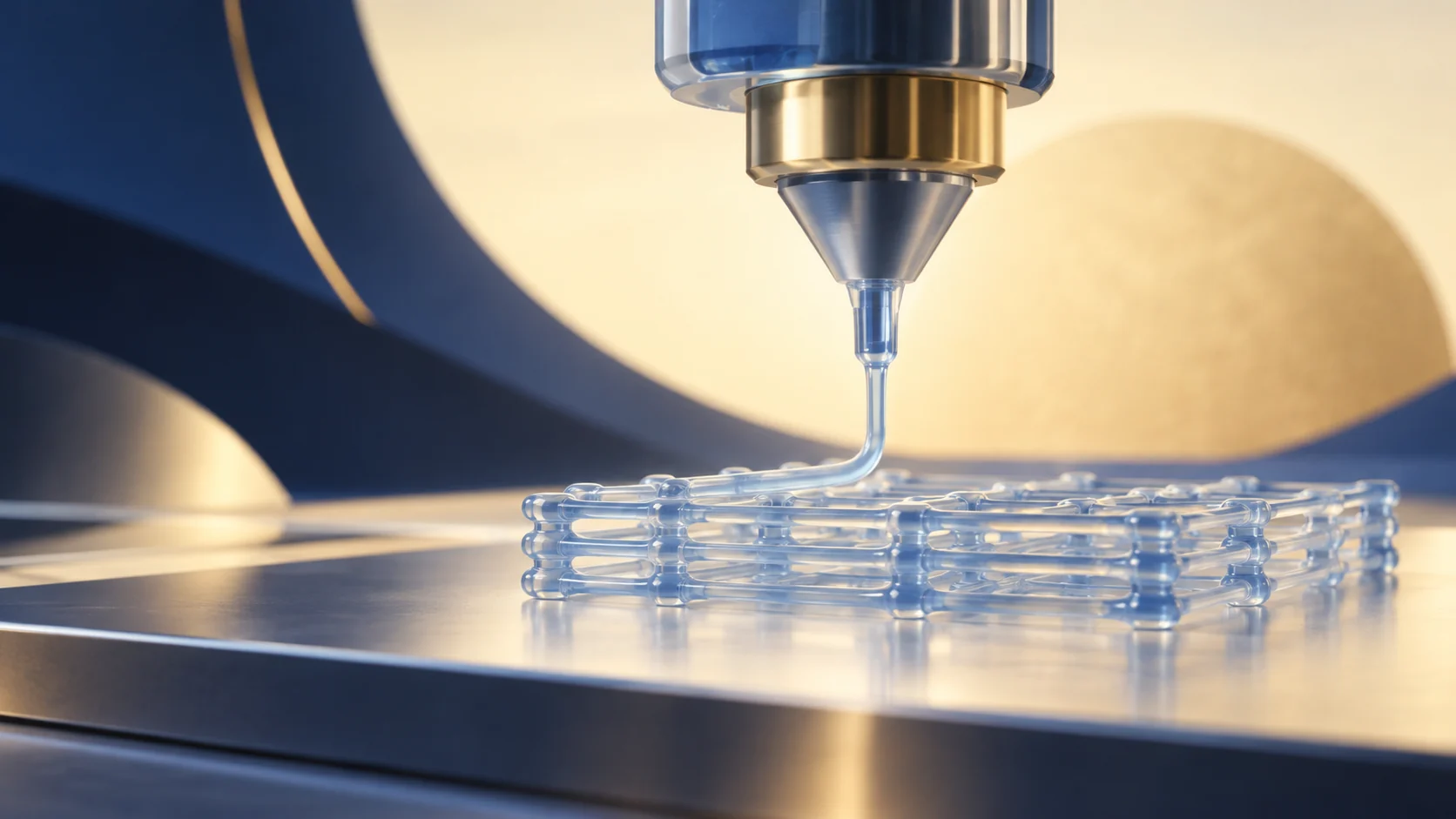
3D bioprinting is a branch of 3D printing proposed to address the limitations of organ transplantation. Conventional transplantation faces limited donor supplies, difficulties with matching and rejection. The emergence of 3D bioprinting offers the possibility of addressing these problems, potentially allowing more patients to receive timely treatment.
How Is a 3D Printing Model Created?
Models for 3D printing can be obtained in several ways. Magnetic resonance imaging (MRI) and computed tomography (CT) can accurately capture the structure of the organ or tissue to be printed. In scientific research, a model can also be drawn using three-dimensional design software such as Solidworks. All 3D models are saved in the standard triangular-mesh format (.stl). The 3D printing software then reads this file and instructs the printer to build the structure layer by layer.
3D Bioprinting
One difference between 3D bioprinting and ordinary 3D printing is that the printing “ink” consists of active components, such as cells and growth factors, mixed with biomaterials. Preparing the ink is a key step in 3D bioprinting. Common biomaterials include hydrogels and decellularized extracellular matrix. Hydrogels contain large amounts of water and are generally biocompatible, making them relatively similar to the environment in the human body; decellularized extracellular matrix provides a more comfortable environment for cell growth. The ink builds living tissue layer by layer. In research designs, printed tissue must first undergo culture and functional testing in vitro. Further validation of safety and efficacy is needed before it can be used for transplantation into humans.
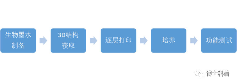
Figure 1: The 3D bioprinting process.
Successful Examples of 3D Bioprinting
In 2017, researchers at Northwestern University in the United States printed overlapping gelatin grids at different angles and placed mouse ovarian follicles within them. They found that blood vessels formed in the follicle-containing scaffolds and that the structures acquired ovarian function. After implantation into mice, the printed structures enabled the animals to give birth to healthy pups.
A leading company in 3D bioprinting is the US company Organovo, which uses its proprietary NovoGen 3D bioprinting technology to print miniature liver structures. Data show that these printed liver structures can produce albumin, cholesterol and detoxification enzymes and metabolize drugs within the liver tissue. Their miniature liver structures have already entered the market, facilitating research on human liver tissue.
3D bioprinting has achieved major research breakthroughs. These findings provide a research foundation for 3D printing tissues and exploring alternatives to organs. Using such technologies in humans to address the shortage of transplantable organs still requires solutions to issues such as functional maturation, vascularization and safety. Meanwhile, 3D printing will also find applications in other areas of the biological sciences, including food, clothing and beauty.
Additional References
The text was recovered from the matching article retained by the WeChat account, with the original site's publication record preserved. The old WeChat promotional layout has been removed, and available original illustrations have been restored.
Editorial note: Restoration and correction on 2026-10-10: corrected the MRI terminology and a typographical error in “ovarian follicles”; checked the mouse ovarian findings against the original 2017 paper. Research tissues are not described as already suitable for routine human transplantation, and no promise is made that this will become possible within a few years.